- denotes a member of the Li Lab
- * equal contribution
- † corresponding author
Key Papers
2026
-
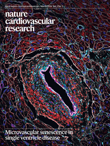 Ventricular assist device unloading reverses microvascular senescence in single ventricle diseaseXiao Li*, Diwakar Turaga*, Yi Zhao, Chang-Ru Tsai, Richard Gang Li, Yuka Morikawa, Hanna J. Tadros, Md Abul Hassan Samee, Iki Adachi, and James F. MartinNature Cardiovascular Research, 2026
Individuals with hypoplastic left heart syndrome (HLHS) have an underdeveloped left ventricle and require surgery to reconfigure blood flow for survival. Here we profiled the HLHS right-ventricular microenvironment by single-nucleus RNA sequencing and spatial transcriptomics at birth (before heart failure), after surgery with heart failure and after ventricular assist device (VAD) unloading (reduced hypoxia and volume overload). We show that HLHS cardiomyocytes, both within the heart and when derived from induced pluripotent stem cells, are intrinsically senescent. The HLHS myocardium contained a senescent microvascular niche with endothelial cells, pericytes and YAP-high fibroblasts, consistent with hypoxic and mechanical stress. This senescent niche is similar to adult myocardial infarction but not pediatric dilated cardiomyopathy with heart failure, pointing to a prominent role of hypoxia in senescence. The microvascular senescent niche was improved by VAD, providing insight into the potential to reverse cardiac cell states that lead to heart failure.
2024
-
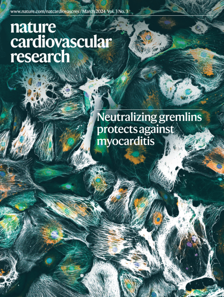 YAP induces a neonatal-like pro-renewal niche in the adult heartRich Gang Li*, Xiao Li*, Yuka Morikawa, Francisco J. Grisanti-Canozo, Fansen Meng, Chang-Ru Tsai, Yi Zhao, Lin Liu, Jong Kim, Bing Xie, Elzbieta Klysik, Shijie Liu, Md Abul Hassan Samee, and James F. MartinNature Cardiovascular Research, 2024
After myocardial infarction (MI), mammalian hearts do not regenerate, and the microenvironment is disrupted. Hippo signaling loss of function with activation of transcriptional co-factor YAP induces heart renewal and rebuilds the post-MI microenvironment. In this study, we investigated adult renewal-competent mouse hearts expressing an active version of YAP, called YAP5SA, in cardiomyocytes (CMs). Spatial transcriptomics and single-cell RNA sequencing revealed a conserved, renewal-competent CM cell state called adult (a)CM2 with high YAP activity. aCM2 co-localized with cardiac fibroblasts (CFs) expressing complement pathway component C3 and macrophages (MPs) expressing C3ar1 receptor to form a cellular triad in YAP5SA hearts and renewal-competent neonatal hearts. Although aCM2 was detected in adult mouse and human hearts, the cellular triad failed to co-localize in these non-renewing hearts. C3 and C3ar1 loss-of-function experiments indicated that C3a signaling between MPs and CFs was required to assemble the pro-renewal aCM2, C3+ CF and C3ar1+ MP cellular triad. Using single-cell RNA sequencing, spatial transcriptomics and genetic experiments, Li et al. report that the close interaction of a specific cardiomyocyte subtype (aCM2), fibroblasts expressing the complement C3 and macrophages expressing C3ar1 was observed in pro-renewal conditions, such as regenerative neonatal hearts and hearts of adult mice overexpressing an active form of YAP in cardiomyocytes.
-
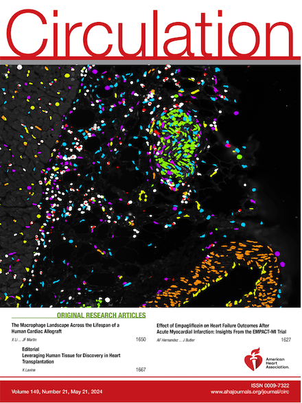 The macrophage landscape across the lifespan of a human cardiac allograftXiao Li*†, Diwakar Turaga*, Rich G Li, Chang-Ru Tsai, Julianna N Quinn, Yi Zhao, Ruby Wilson, Katherine Carlson, Jun Wang, Joseph A Spinner, Edward J Hickey, Iki Adachi, and James F Martin†Circulation, 2024
BACKGROUND: Much of our knowledge of organ rejection after transplantation is derived from rodent models. METHODS: We used single-nucleus RNA sequencing to investigate the inflammatory myocardial microenvironment in human pediatric cardiac allografts at different stages after transplantation. We distinguished donor- from recipient-derived cells using naturally occurring genetic variants embedded in single-nucleus RNA sequencing data. RESULTS: Donor-derived tissue resident macrophages, which accompany the allograft into the recipient, are lost over time after transplantation. In contrast, monocyte-derived macrophages from the recipient populate the heart within days after transplantation and form 2 macrophage populations: recipient MP1 and recipient MP2. Recipient MP2s have cell signatures similar to donor-derived resident macrophages; however, they lack signatures of pro-reparative phagocytic activity typical of donor-derived resident macrophages and instead express profibrotic genes. In contrast, recipient MP1s express genes consistent with hallmarks of cellular rejection. Our data suggest that recipient MP1s activate a subset of natural killer cells, turning them into a cytotoxic cell population through feed-forward signaling between recipient MP1s and natural killer. CONCLUSIONS: Our findings reveal an imbalance of donor-derived and recipient-derived macrophages in the pediatric cardiac allograft that contributes to allograft failure.
2023
-
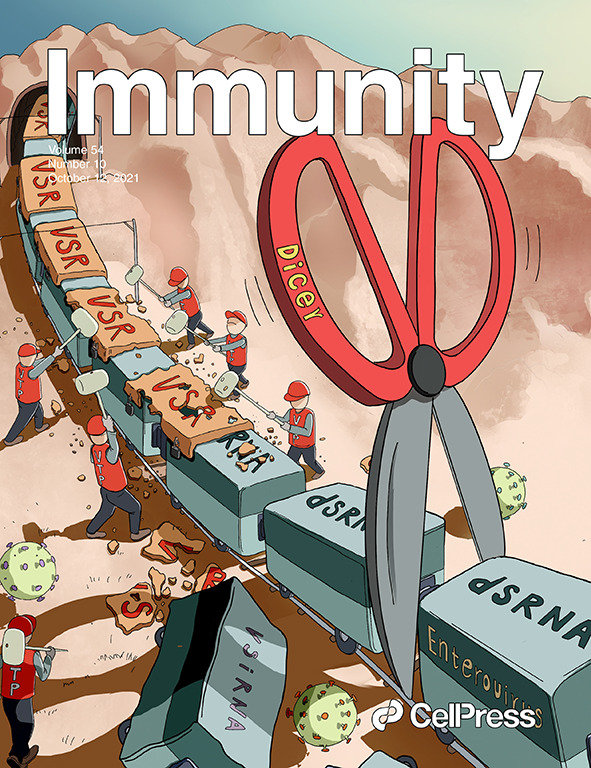
 Lymphatic endothelial transcription factor Tbx1 promotes an immunosuppressive microenvironment to facilitate post-myocardial infarction repairWenfeng Wang*, Xiao Li*, Xiaoning Ding*, Shanshan Xiong*, Zhenlei Hu, Xuan Lu, Kan Zhang, Heng Zhang, Qianwen Hu, Kaa Seng Lai, Zhongxiang Chen, Junjie Yang, Hejie Song, Ye Wang, Lu Wei, Zeyang Xia, Bin Zhou, Yulong He, Jun Pu, Xiao Liu, Rongqin Ke, Tao Wu, Chuanxin Huang, Antonio Baldini, Min Zhang, and Zhen ZhangImmunity, 2023
The heart is an autoimmune-prone organ. It is crucial for the heart to keep injury-induced autoimmunity in check to avoid autoimmune-mediated inflammatory disease. However, little is known about how injury-induced autoimmunity is constrained in hearts. Here, we reveal an unknown intramyocardial immunosuppressive program driven by Tbx1, a DiGeorge syndrome disease gene that encodes a T-box transcription factor (TF). We found induced profound lymphangiogenic and immunomodulatory gene expression changes in lymphatic endothelial cells (LECs) after myocardial infarction (MI). The activated LECs penetrated the infarcted area and functioned as intramyocardial immune hubs to increase the numbers of tolerogenic dendritic cells (tDCs) and regulatory T (Treg) cells through the chemokine Ccl21 and integrin Icam1, thereby inhibiting the expansion of autoreactive CD8+ T cells and promoting reparative macrophage expansion to facilitate post-MI repair. Mimicking its timing and implementation may be an additional approach to treating autoimmunity-mediated cardiac diseases.
Editorial Highlights- Elisa Martini (2023). Lymphatic endothelial cells orchestrate cardiac immunotolerance after myocardial infarction. Nature Cardiovascular Research.
- Yvonne Bordon (2023). Lymphatic expression of TBX1 helps to heal the heart. Nature Reviews Immunology.
- Sara Perrotta and Daniela Carnevale (2023). Tbx1 orchestrates an immune niche that safeguards a broken heart. Immunity.
2021
-
 Distinct human Langerhans cell subsets orchestrate reciprocal functions and require different developmental regulationXiaochun Liu, Ronghui Zhu, Yang Luo, Shangshang Wang, Yi Zhao, Zhuoqiong Qiu, Yu Zhang, Xiao Liu, Xu Yao†, Xiao Li†, and Wei Li†Immunity, 2021
Langerhans cells (LCs) play a pivotal role in skin homeostasis, and the heterogeneity of LCs has long been considered. In this study, we have identified two steady-state (LC1 and LC2) and two activated LC subsets in the epidermis of human skin and in LCs derived from CD34+ hemopoietic stem cells (HSC-LCs) by utilizing single-cell RNA sequencing and mass cytometry. Analysis of HSC-LCs at multiple time-points during differentiation revealed that EGR1 and Notch signaling were among the top pathways regulating the bifurcation of LC1 and LC2. LC1 were characterized as classical LCs, mainly related to innate immunity and antigen processing. LC2 were similar to monocytes or myeloid dendritic cells, involving in immune responses and leukocyte activation. LC1 remained stable under inflammatory microenvironment, whereas LC2 were prone to being activated and demonstrated elevated expression of immuno-suppressive molecules. We revealed distinct human LC subsets that require different developmental regulation and orchestrate reciprocal functions.
2019
-
 Chromatin-associated RNAs as facilitators of functional genomic interactionsXiao Li and Xiang-Dong FuNature Reviews Genetics, Jun 2019
Mammalian genomes are extensively transcribed, which produces a large number of both coding and non-coding transcripts. Various RNAs are physically associated with chromatin, through being either retained in cis at their site of transcription or recruited in trans to other genomic regions. Driven by recent technological innovations for detecting chromatin-associated RNAs, diverse roles are being revealed for these RNAs and associated RNA-binding proteins (RBPs) in gene regulation and genome function. Such functions include locus-specific roles in gene activation and silencing, as well as emerging roles in higher-order genome organization, such as involvement in long-range enhancer–promoter interactions, transcription hubs, heterochromatin, nuclear bodies and phase transitions. Enabled by genome-wide profiling approaches, there is growing appreciation for the prevalence and functional importance of various types of chromatin-associated RNAs. As Li and Fu describe in this Review, these RNAs can either be retained in cis at their site of transcription or recruited in cis to other loci, and they have diverse roles in gene regulation, genome organization, nuclear body formation and phase-separation events.
2017
-
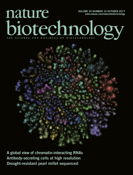 GRID-seq reveals the global RNA–chromatin interactomeXiao Li*, Bing Zhou*, Liang Chen, Lan-Tao Gou, Hairi Li, and Xiang-Dong FuNature Biotechnology, 2017
The RNAs bound to the genome and their binding sites are detected with GRID-seq. Higher eukaryotic genomes are bound by a large number of coding and non-coding RNAs, but approaches to comprehensively map the identity and binding sites of these RNAs are lacking. Here we report a method to capture in situ global RNA interactions with DNA by deep sequencing (GRID-seq), which enables the comprehensive identification of the entire repertoire of chromatin-interacting RNAs and their respective binding sites. In human, mouse, and Drosophila cells, we detected a large set of tissue-specific coding and non-coding RNAs that are bound to active promoters and enhancers, especially super-enhancers. Assuming that most mRNA–chromatin interactions indicate the physical proximity of a promoter and an enhancer, we constructed a three-dimensional global connectivity map of promoters and enhancers, revealing transcription-activity-linked genomic interactions in the nucleus.
Publications in the Past 10 Years
2026
- YAP induces a prorenewal metabolic state in cardiomyocytesLin Liu, Jeffrey D. Steimle, Chang-Ru Tsai, Fansen Meng, Yuka Morikawa, Yi Zhao, Sandra Carmichael, Xiao Li, and James F. MartinCirculation, 2026
Cardiomyocytes, as highly specialized and differentiated somatic cells, possess a limited capacity for renewal. Neonatal rodents possess the ability to regenerate cardiomyocytes after injury; however, this regenerative capacity declines rapidly with cardiomyocyte maturation, suggesting an inhibitory network between cellular maturation and cardiomyocyte proliferation. Maturing cardiomyocytes undergo a metabolic shift from predominantly glycolysis in the neonatal state to increased fatty acid oxidation in the mature state, which poses a barrier to cardiomyocyte proliferation and cardiac regenerative repair. YAP, a transcriptional cofactor regulated by the Hippo signaling pathway, promotes cardiac regenerative repair. We investigated the role of YAP in mediating metabolic remodeling to overcome the cardiomyocyte proliferation barrier and enable cardiac regenerative repair after heart injury. We explored how YAP induces metabolic remodeling through single-nucleus RNA sequencing and metabolomic analyses in mice. Using lipidomic analysis, we demonstrated how YAP remodels the balance of fatty acid catabolism and anabolism. We further used a maternal fat overloading model to stimulate fatty acid oxidation, which activates a maturation program in neonatal cardiomyocytes and counteracts YAP-mediated metabolic dematuration. Using chromatin accessibility (assay for transposase-accessible chromatin with high-throughput sequencing), DNA footprinting, and transcriptional profiling (RNA sequencing), we discovered the key transcription factors that YAP interrupts to reprogram the cardiomyocyte metabolic state. Our results demonstrate that YAP directs metabolic remodeling of mature cardiomyocytes toward a neonatal-like metabolic state and illustrate the role of fatty acid metabolism in proliferating cardiomyocytes. We found that YAP reduces cardiomyocyte fatty acid utilization, driving fatty acid anabolism and phospholipid biosynthesis. Genome-wide analyses revealed that YAP inhibits the cardiac maturation transcription factor MEF2A (myocyte-specific enhancer factor 2A), resulting in decreased gene expression of cardiomyocyte maturity pathways. Given the role of MEF2A in regulating contractility, energy production, and mitochondrial homeostasis, we found that perturbing MEF2A transcriptional activity can serve as a strategy to interrupt the cardiomyocyte maturation program and restore the regenerative capacity of the heart. Our research endeavors to provide a comprehensive understanding of the balance of cardiomyocyte metabolic maturation and proliferation to overcome barriers to heart regeneration, offering novel insights into the potential for therapeutic intervention in heart failure.
- Ventricular assist device unloading reverses microvascular senescence in single ventricle diseaseXiao Li*, Diwakar Turaga*, Yi Zhao, Chang-Ru Tsai, Richard Gang Li, Yuka Morikawa, Hanna J. Tadros, Md Abul Hassan Samee, Iki Adachi, and James F. MartinNature Cardiovascular Research, 2026
Individuals with hypoplastic left heart syndrome (HLHS) have an underdeveloped left ventricle and require surgery to reconfigure blood flow for survival. Here we profiled the HLHS right-ventricular microenvironment by single-nucleus RNA sequencing and spatial transcriptomics at birth (before heart failure), after surgery with heart failure and after ventricular assist device (VAD) unloading (reduced hypoxia and volume overload). We show that HLHS cardiomyocytes, both within the heart and when derived from induced pluripotent stem cells, are intrinsically senescent. The HLHS myocardium contained a senescent microvascular niche with endothelial cells, pericytes and YAP-high fibroblasts, consistent with hypoxic and mechanical stress. This senescent niche is similar to adult myocardial infarction but not pediatric dilated cardiomyopathy with heart failure, pointing to a prominent role of hypoxia in senescence. The microvascular senescent niche was improved by VAD, providing insight into the potential to reverse cardiac cell states that lead to heart failure.
- Single-nuclei transcriptomic profiling of human myocardium in long-chain 3-hydroxyacyl-coenzyme A dehydrogenase deficiencyHanna J. Tadros, Diwakar Turaga, Yi Zhao, Chang-Ru Tsai, Lalita Wadhwa, Debra L. Kearney, Iki Adachi, Xiao Li, and James F. MartinCirculation: Heart Failure, 2026
- Single cell transcriptomic landscape of adult and pediatric non-ischemic cardiomyopathyHanna J. Tadros, Diwakar Turaga, Kyle Hope, Joseph A. Spinner, Iki Adachi, Xiao Li, and James F. MartinJournal of Molecular and Cellular Cardiology, 2026
Non-ischemic cardiomyopathy (NICM) is a devastating diagnosis with a wide array of phenotypes, ranging from mild cardiac hypertrophy to end-stage heart failure. Single-cell/nucleus RNA sequencing technologies have expanded and become a necessary tool to unravel the transcriptome across thousands to millions of cells. Studies incorporating these technologies to examine pediatric and adult myocardium have improved our understanding of underlying pathophysiology in cardiomyopathy, identified novel gene and genetic pathway associations, and paved the way for precision medicine therapeutics in cardiovascular medicine. We compiled the recent literature that showcase single cell/nucleus technologies in NICM in adult and pediatric populations and describe cell type-specific changes, ultimately setting the stage for future targeted gene manipulation/precision medicine.
- Microvascular senescence in single ventricle diseaseXiao Li and James F. MartinNature Cardiovascular Research, 2026
We performed single-nucleus and spatial transcriptomic analyses of systemic right ventricles from pediatric donors with hypoplastic left heart syndrome after surgical palliation. Our analyses demonstrate widespread senescence in the failing heart, spatially organized into microvascular niches, and show that these pathological states are partially reversible with mechanical unloading.
2025
- YAP-induced glycolysis drives fibroinflammation and disrupts fibroblast fidelityChang-Ru Tsai, Lin Liu, Yi Zhao, Jong H. Kim, Paulo Czarnewski, Rich Gang Li, Fansen Meng, Mingjie Zheng, Jeffrey Steimle, Xiaolei Zhao, Francisco Grisanti, Zheng Sun, Jun Wang, Md. Abul Hassan Samee, Xiao Li, and James F. MartinCirculation Research, 2025
Separation of the pulmonic and systemic circulation is essential for terrestrial life, and mammals have evolved distinct cardiac chambers with specialized structures and functions. Transcriptomics profiling revealed cellular heterogeneity between heart chambers. However, the mechanisms underlying chamber-specific transcriptomic and metabolic differences—and their functional significance—remain poorly understood. The Hippo/YAP (yes-associated protein) pathway is a conserved signaling network that regulates diverse cellular processes. The Hippo kinases inhibit YAP in cardiac fibroblasts (CF) to restrict fibrosis and inflammation. Nonetheless, how YAP regulates the metabolic microenvironment during homeostasis and fibroinflammation remains unclear. We investigated YAP and glycolysis activity in the 4 cardiac chambers by scoring the expression of YAP target genes and glycolysis genes in human single-nucleus RNA sequencing data. To compare glucose uptake between the left and right atria, we measured isotope-labeled glucose uptake in isolated mouse atria. To study the role of YAP in CFs, we inactivated the Hippo kinases, Lats1 and Lats2, in mouse CFs and performed metabolic studies, snRNA-seq, single-nucleus assay for transposase-accessible chromatin with sequencing, and spatial transcriptomics. Metabolic and sequencing approaches revealed that Hippo-deficient CFs activated glycolysis to promote fibroinflammation. Inhibition of glycolysis or lactate production suppressed Hippo-deficient CF-induced fibrosis. Elevated YAP activity disrupted fibroblast lineage fidelity by inducing an osteochondroprogenitor cell state. Blocking macrophage expansion pharmacologically reduced Hippo-deficient CF proliferation and fibrosis. Sequencing and functional studies showed that macrophages secreted IGF1 (insulin-like growth factor 1) to activate IGF1 signaling in Hippo-deficient CFs to increase cell proliferation and fibrosis. We discovered that right atrial CFs are more glycolytic and have higher YAP activity than CFs in other heart chambers. YAP activation in CFs induces glycolysis to drive fibrosis. YAP disrupts fibroblast lineage fidelity, driving them to a SOX9 (SRY-box transcription factor 9)-expressing osteochondroprogenitor cell state. Mechanistically, YAP activates the secretion of CSF1 (colony-stimulating factor 1) to promote macrophage expansion. Blocking macrophage expansion reduces Hippo-deficient CF proliferation, osteochondroprogenitor differentiation, and fibrosis, revealing that macrophages signal reciprocally to regulate CF cell states. Genomic and functional studies revealed that the upregulated IGF1 receptor in Hippo-deficient CFs enables them to receive macrophage-secreted IGF1, thereby further enhancing CF proliferation and fibrosis.
- Single-nuclei multiomics analysis identifies abnormal cardiomyocytes in a murine model of cardiac developmentRiley Leonard, Yi Zhao, Steven Eliason, Kathy Zimmerman, Ariana Batz, Cathy J. Hatcher, Robert M. Weiss, Mason Sweat, Xiao Li, and Brad A. AmendtNature Communications, 2025
Transcription factors such as Tbx5, Gata4, Mef2c and Pitx2 are required during cardiac development, and in adult cardiac homeostasis. We demonstrate that the gene dosage and modulation of these factors are mediated in vivo by the miR-200 family. Inhibition of a single miR-200 family member within the cluster results in defects of the left ventricle and cardiomyocyte maturation during development. Inhibition of the entire miR-200 family results in a ventricular septal defect and embryonic lethality by embryonic day (E)16.5. Inhibition of each miR-200 family has distinct heart phenotypes in cell specific differentiation and maturation. snRNA-sequencing reveals an immature cardiomyocyte cell state, suggesting reduced differentiation of these cells. The miR-200 family members are critical regulators of early cardiac development through maintaining cardiomyocyte differentiation and maturation. In this report, we identify several transcription factors regulated by miR-200 during heart development, a role for miR-200 in specific heart defects, and an abnormal cardiomyocyte population.
- SARS-CoV-2 NSP13 interacts with TEAD to suppress Hippo-YAP signalingFansen Meng, Jong Hwan Kim, Chang-Ru Tsai, Jeffrey D Steimle, Jun Wang, Yufeng Shi, Rich G Li, Bing Xie, Vaibhav Deshmukh, Shijie Liu, Xiao Li, and James F MartineLife, 2025
The Hippo pathway controls organ development, homeostasis, and regeneration primarily by modulating YAP/TEAD-mediated gene expression. Although emerging studies report Hippo-YAP dysfunction after viral infection, it is largely unknown in the context of severe acute respiratory syndrome coronavirus 2 (SARS-CoV-2). Here, we analyzed RNA sequencing data from human-induced pluripotent stem cell-derived cardiomyocytes (iPSC-CMs) and SARS-CoV-2-infected human lung samples, and observed a decrease in YAP target gene expression. In screening SARS-CoV-2 nonstructural proteins, we found that nonstructural protein 13 (NSP13), a conserved coronavirus helicase, inhibits YAP transcriptional activity independent of the upstream Hippo kinases LATS1/2. Consistently, introducing NSP13 into mouse cardiomyocytes suppresses an active form of YAP (YAP5SA) in vivo . Subsequent investigations on NSP13 mutants revealed that NSP13 helicase activity, including DNA binding and unwinding, is crucial for suppressing YAP transactivation in HEK293T cells. Mechanistically, TEAD4 serves as a platform to recruit NSP13 and YAP. NSP13 likely inactivates the YAP/TEAD4 transcription complex by remodeling chromatin to recruit proteins, such as transcription termination factor 2 (TTF2), to bind the YAP/TEAD/NSP13 complex. These findings reveal a novel YAP/TEAD regulatory mechanism and uncover molecular insights into Hippo-YAP regulation after SARS-CoV-2 infection in humans.
- Loss of methyltransferase and hypomethylated m6A sarcomere transcripts leading to early-onset dilated cardiomyopathySophie Marie Hand, Yi Zhao, Donna Li, Savanna Burke, Hee-Woong Lim, Jessie Yester, Vidu Garg, Federica Accornero, Xiao Li, Deqiang Li, and Jihyun JangCirculation, 2025
- Gene therapy CM-YAPon protects the mouse heart from myocardial infarctionFansen Meng, Jeffrey D. Steimle, Elizabeth Straight, Rich G. Li, Yuka Morikawa, Zohaib Iqbal, Bing Xie, Jun Wang, Wyatt G. Paltzer, Yi Zhao, Chang-Ru Tsai, Lin Liu, Maggie Lim, Rita A. Schack, Daniel Ramirez, Katherine Carlson, Vaibhav Deshmukh, Jason M. Karch, Robia G. Pautler, Xiao Li, and James F. MartinNature Cardiovascular Research, 2025
Myocardial infarction (MI) affects millions of people worldwide, causing irreversible injury to the heart and impairing cardiac function1. In both mouse and pig MI models, activating YAP in cardiomyocytes (CMs) stimulates regenerative repair2,3. Here we develop an adeno-associated virus 9-based therapy, termed CM-YAPon, which enables transient expression of an active YAP variant (YAP5SA) in CMs after exposure to the small molecule LMI070. A single LMI070 dose in mice triggers YAP5SA expression, CM cell cycle reentry and reprogramming of the cardiac microenvironment. YAP5SA induction after MI rapidly improves cardiac function while pre-MI induction confers cardioprotection and reduces cell death across multiple cardiac cell types. These findings reveal the therapeutic potential of reversible gene activation for ischemic heart disease.
- Fibroblasts are the primary contributors to a disrupted micro-environment in end-stage pediatric hypertrophic cardiomyopathyHanna J Tadros, Diwakar Turaga, Yi Zhao, Chang-Ru Tsai, Iki A Adachi, Xiao Li, and James F MartinCirculation: Genomic and Precision Medicine, 2025
BACKGROUND: Hypertrophic cardiomyopathy (HCM) is a relatively rare but debilitating diagnosis in the pediatric population, and patients with end-stage HCM require heart transplantation. Here, we have examined the transcriptome in ventricular tissue from this patient group to identify cell states and underlying cellular processes unique to pediatric HCM. METHODS: We performed single-nucleus RNA sequencing on explanted hearts at transplant in 3 pediatric patients with end-stage HCM and compared findings to pediatric control and adult HCM. RESULTS: We identified distinct underlying cellular processes in cardiomyocytes, fibroblasts, endothelial cells, and myeloid cells compared with controls. Pediatric HCM was enriched in cardiomyocytes exhibiting stressed myocardium gene signatures and underlying pathways associated with cardiac hypertrophy; cardiac fibroblasts exhibited activation signatures and compared with adult patients, exhibited heightened downstream processes associated with fibrosis and a unique, myofibroblast-like cluster with increased metabolic stress and antiapoptotic properties. We noted depletion of tissue-resident macrophages and increased vascular remodeling in endothelial cells in pediatric HCM. CONCLUSIONS: Our analysis provides the first single-nucleus analysis focused on end-stage pediatric HCM. Fibroblast-mediated cellular processes were the most prominent in pediatric HCM, which had more downstream processes associated with fibrosis than did adult HCM.
2024
- Activated fibroblasts drive cellular interactions in end-stage pediatric hypertrophic cardiomyopathyHanna J. Tadros, Diwakar Turaga, Yi Zhao, Chang-Ru Tsai, Iki A. Adachi, Xiao Li, and James F. MartinbioRxiv, 2024
Hypertrophic cardiomyopathy (HCM) is a relatively rare but debilitating diagnosis in the pediatric population and patients with end-stage HCM require heart transplantation. In this study, we performed single-nucleus RNA sequencing on pediatric HCM and control myocardium. We identified distinct underling cellular processes in pediatric, end-stage HCM in cardiomyocytes, fibroblasts, endothelial cells, and myeloid cells, compared to controls. Pediatric HCM was enriched in cardiomyocytes exhibiting “stressed” myocardium gene signatures and underlying pathways associated with cardiac hypertrophy. Cardiac fibroblasts exhibited clear activation signatures and heightened downstream processes associated with fibrosis, more so than adult counterparts. There was notable depletion of tissue-resident macrophages, and increased vascular remodeling in endothelial cells. Our analysis provides the first single nuclei analysis focused on end-stage pediatric HCM.
- YAP induces a neonatal-like pro-renewal niche in the adult heartRich Gang Li*, Xiao Li*, Yuka Morikawa, Francisco J. Grisanti-Canozo, Fansen Meng, Chang-Ru Tsai, Yi Zhao, Lin Liu, Jong Kim, Bing Xie, Elzbieta Klysik, Shijie Liu, Md Abul Hassan Samee, and James F. MartinNature Cardiovascular Research, 2024
After myocardial infarction (MI), mammalian hearts do not regenerate, and the microenvironment is disrupted. Hippo signaling loss of function with activation of transcriptional co-factor YAP induces heart renewal and rebuilds the post-MI microenvironment. In this study, we investigated adult renewal-competent mouse hearts expressing an active version of YAP, called YAP5SA, in cardiomyocytes (CMs). Spatial transcriptomics and single-cell RNA sequencing revealed a conserved, renewal-competent CM cell state called adult (a)CM2 with high YAP activity. aCM2 co-localized with cardiac fibroblasts (CFs) expressing complement pathway component C3 and macrophages (MPs) expressing C3ar1 receptor to form a cellular triad in YAP5SA hearts and renewal-competent neonatal hearts. Although aCM2 was detected in adult mouse and human hearts, the cellular triad failed to co-localize in these non-renewing hearts. C3 and C3ar1 loss-of-function experiments indicated that C3a signaling between MPs and CFs was required to assemble the pro-renewal aCM2, C3+ CF and C3ar1+ MP cellular triad. Using single-cell RNA sequencing, spatial transcriptomics and genetic experiments, Li et al. report that the close interaction of a specific cardiomyocyte subtype (aCM2), fibroblasts expressing the complement C3 and macrophages expressing C3ar1 was observed in pro-renewal conditions, such as regenerative neonatal hearts and hearts of adult mice overexpressing an active form of YAP in cardiomyocytes.
- The macrophage landscape across the lifespan of a human cardiac allograftXiao Li*†, Diwakar Turaga*, Rich G Li, Chang-Ru Tsai, Julianna N Quinn, Yi Zhao, Ruby Wilson, Katherine Carlson, Jun Wang, Joseph A Spinner, Edward J Hickey, Iki Adachi, and James F Martin†Circulation, 2024
BACKGROUND: Much of our knowledge of organ rejection after transplantation is derived from rodent models. METHODS: We used single-nucleus RNA sequencing to investigate the inflammatory myocardial microenvironment in human pediatric cardiac allografts at different stages after transplantation. We distinguished donor- from recipient-derived cells using naturally occurring genetic variants embedded in single-nucleus RNA sequencing data. RESULTS: Donor-derived tissue resident macrophages, which accompany the allograft into the recipient, are lost over time after transplantation. In contrast, monocyte-derived macrophages from the recipient populate the heart within days after transplantation and form 2 macrophage populations: recipient MP1 and recipient MP2. Recipient MP2s have cell signatures similar to donor-derived resident macrophages; however, they lack signatures of pro-reparative phagocytic activity typical of donor-derived resident macrophages and instead express profibrotic genes. In contrast, recipient MP1s express genes consistent with hallmarks of cellular rejection. Our data suggest that recipient MP1s activate a subset of natural killer cells, turning them into a cytotoxic cell population through feed-forward signaling between recipient MP1s and natural killer. CONCLUSIONS: Our findings reveal an imbalance of donor-derived and recipient-derived macrophages in the pediatric cardiac allograft that contributes to allograft failure.
- Single-nuclei transcriptomics reveals TBX5-dependent targets in a patient with Holt-Oram syndromeJeffrey D. Steimle, Yi Zhao, Fansen Meng, Mikaela E. Taylor, Diwakar Turaga, Iki Adachi, Xiao Li, and James F. MartinJournal of Clinical Investigation, 2024
- Single nucleus transcriptome of a “Super RV” shows increased insulin and angiogenesis signalingDiwakar Turaga*, Xiao Li*, Yi Zhao, Chang-Ru Tsai, Axel Moreira, Edward Hickey, Iki Adachi, and James MartinbioRxiv, 2024
The right ventricle (RV) is one of the four pumping chambers of the heart, pumping blood to the lungs. In severe forms of congenital heart disease and pulmonary hypertension, the RV is made to pump into the systemic circulation. Such systemic RVs typically display early failure due to pressure overload. In rare cases a systemic RV persists into later decades of life – colloquially called a ‘Super RV’. Here we present the single-nucleus transcriptome of a systemic RV from a 60-year-old with congenitally corrected transposition of great arteries (ccTGA). Our data shows two specific signaling pathways enriched in the ccTGA RV myocardium. First, we show increased insulin like growth factor (IGF1) signaling within the systemic RV myocardium: there is increased expression of the main receptor IGFR1 within the cardiomyocytes, and IGF1 ligands within the cardiofibroblasts and macrophages. Second, we find increased VEGF and Wnt9 ligand expression in cardiomyocytes and increased VEGF1R and Wnt9 receptors in endothelial cells, which are implicated in angiogenesis. We show that increased insulin and angiogenesis signaling are potentially beneficial RV adaptations to increased pressure overload. This study of an adult systemic RV provides an important framework for understanding RV remodeling to systemic pressures in congenital heart disease and pulmonary hypertension.
- Myocardial infarction suppresses protein synthesis and causes decoupling of transcription and translationShijie Liu, Vaibhav Deshmukh, Fangfei Wang, Jie Liang, Jenna Cusick, Xiao Li, and James F. MartinJACC: Basic to Translational Science, 2024
Gene expression involves transcription, translation, and mRNA and protein degradation. Advanced RNA sequencing measures mRNA levels for cell state assessment, but mRNA level does not fully reflect protein level. Identifying heart cell proteomes and their stress response is crucial. Using a cardiomyocyte-specific mouse model, we tracked protein synthesis after myocardial infarction. Our results showed that myocardial infarction suppresses protein synthesis and unveils a decoupling of translation and transcription regulation in cardiomyocytes.
- Endothelial cells adopt a pro-reparative immune responsive signature during cardiac injuryHali Long, Jeffrey D Steimle, Francisco Jose Grisanti Canozo, Jong Hwan Kim, Xiao Li, Yuka Morikawa, Minjun Park, Diwakar Turaga, Iki Adachi, Joshua D Wythe, Md Abul Hassan Samee, and James F MartinLife Science Alliance, 2024
Modulation of the heart’s immune microenvironment is crucial for recovery after ischemic events such as myocardial infarction (MI). Endothelial cells (ECs) can have immune regulatory functions; however, interactions between ECs and the immune environment in the heart after MI remain poorly understood. We identified an EC-specific IFN responsive and immune regulatory gene signature in adult and pediatric heart failure (HF) tissues. Single-cell transcriptomic analysis of murine hearts subjected to MI uncovered an EC population (IFN-ECs) with immunologic gene signatures similar to those in human HF. IFN-ECs were enriched in regenerative-stage mouse hearts and expressed genes encoding immune responsive transcription factors (Irf7, Batf2, and Stat1). Single-cell chromatin accessibility studies revealed an enrichment of these TF motifs at IFN-EC signature genes. Expression of immune regulatory ligand genes by IFN-ECs suggests bidirectional signaling between IFN-ECs and macrophages in regenerative-stage hearts. Our data suggest that ECs may adopt immune regulatory signatures after cardiac injury to accompany the reparative response. The presence of these signatures in human HF and murine MI models suggests a potential role for EC-mediated immune regulation in responding to stress induced by acute injury in MI and chronic adverse remodeling in HF.
2023
- Hippo-deficient cardiac fibroblasts differentiate into osteochondroprogenitorsChang-Ru Tsai, Jong Kim, Xiao Li, Paulo Czarnewski, Rich Li, Fansen Meng, Mingjie Zheng, Xiaolei Zhao, Jeffrey Steimle, Francisco Grisanti, Jun Wang, Md. Abul Hassan Samee, and James MartinbioRxiv, 2023
Cardiac fibrosis, a common pathophysiology associated with various heart diseases, occurs from the excess deposition of extracellular matrix (ECM)1. Cardiac fibroblasts (CFs) are the primary cells that produce, degrade, and remodel ECM during homeostasis and tissue repair2. Upon injury, CFs gain plasticity to differentiate into myofibroblasts3 and adipocyte-like4,5 and osteoblast-like6 cells, promoting fibrosis and impairing heart function7. How CFs maintain their cell state during homeostasis and adapt plasticity upon injury are not well defined. Recent studies have shown that Hippo signalling in CFs regulates cardiac fibrosis and inflammation8–11. Here, we used single-nucleus RNA sequencing (snRNA-seq) and spatially resolved transcriptomic profiling (ST) to investigate how the cell state was altered in the absence of Hippo signaling and how Hippo-deficient CFs interact with macrophages during cardiac fibrosis. We found that Hippo-deficient CFs differentiate into osteochondroprogenitors (OCPs), suggesting that Hippo restricts CF plasticity. Furthermore, Hippo-deficient CFs colocalized with macrophages, suggesting their intercellular communications. Indeed, we identified several ligand-receptor pairs between the Hippo-deficient CFs and macrophages. Blocking the Hippo-deficient CF-induced CSF1 signaling abolished macrophage expansion. Interestingly, blocking macrophage expansion also reduced OCP differentiation of Hippo-deficient CFs, indicating that macrophages promote CF plasticity.
- Response to identifying the epidermal dendritic cell landscapeRonghui Zhu, Xiaochun Liu, Xiao Li†, Xu Yao†, and Wei Li†Immunity, 2023
- Lymphatic endothelial transcription factor Tbx1 promotes an immunosuppressive microenvironment to facilitate post-myocardial infarction repairWenfeng Wang*, Xiao Li*, Xiaoning Ding*, Shanshan Xiong*, Zhenlei Hu, Xuan Lu, Kan Zhang, Heng Zhang, Qianwen Hu, Kaa Seng Lai, Zhongxiang Chen, Junjie Yang, Hejie Song, Ye Wang, Lu Wei, Zeyang Xia, Bin Zhou, Yulong He, Jun Pu, Xiao Liu, Rongqin Ke, Tao Wu, Chuanxin Huang, Antonio Baldini, Min Zhang, and Zhen ZhangImmunity, 2023
The heart is an autoimmune-prone organ. It is crucial for the heart to keep injury-induced autoimmunity in check to avoid autoimmune-mediated inflammatory disease. However, little is known about how injury-induced autoimmunity is constrained in hearts. Here, we reveal an unknown intramyocardial immunosuppressive program driven by Tbx1, a DiGeorge syndrome disease gene that encodes a T-box transcription factor (TF). We found induced profound lymphangiogenic and immunomodulatory gene expression changes in lymphatic endothelial cells (LECs) after myocardial infarction (MI). The activated LECs penetrated the infarcted area and functioned as intramyocardial immune hubs to increase the numbers of tolerogenic dendritic cells (tDCs) and regulatory T (Treg) cells through the chemokine Ccl21 and integrin Icam1, thereby inhibiting the expansion of autoreactive CD8+ T cells and promoting reparative macrophage expansion to facilitate post-MI repair. Mimicking its timing and implementation may be an additional approach to treating autoimmunity-mediated cardiac diseases.
Editorial Highlights- Elisa Martini (2023). Lymphatic endothelial cells orchestrate cardiac immunotolerance after myocardial infarction. Nature Cardiovascular Research.
- Yvonne Bordon (2023). Lymphatic expression of TBX1 helps to heal the heart. Nature Reviews Immunology.
- Sara Perrotta and Daniela Carnevale (2023). Tbx1 orchestrates an immune niche that safeguards a broken heart. Immunity.
2022
- HMGN2 represses gene transcription via interaction with transcription factors Lef-1 and Pitx2 during amelogenesisSteven Eliason, Dan Su, Flavia Pinho, Zhao Sun, Zichao Zhang, Xiao Li, Mason Sweat, Shankar R. Venugopalan, Bing He, Michael Bustin, and Brad A. AmendtJournal of Biological Chemistry, 2022
The chromatin-associated high mobility group protein N2 (HMGN2) cofactor regulates transcription factor activity through both chromatin and protein interactions. Hmgn2 expression is known to be developmentally regulated, but the post-transcriptional mechanisms that regulate Hmgn2 expression and its precise roles in tooth development remain unclear. Here, we demonstrate that HMGN2 inhibits the activity of multiple transcription factors as a general mechanism to regulate early development. Bimolecular fluorescence complementation, pull-down, and coimmunoprecipitation assays show that HMGN2 interacts with the transcription factor Lef-1 through its HMG-box domain as well as with other early development transcription factors, Dlx2, FoxJ1, and Pitx2. Furthermore, EMSAs demonstrate that HMGN2 binding to Lef-1 inhibits its DNA-binding activity. We found that Pitx2 and Hmgn2 associate with H4K5ac and H3K4me2 chromatin marks in the proximal Dlx2 promoter, demonstrating Hmgn2 association with open chromatin. In addition, we demonstrate that microRNAs (miRs) mir-23a and miR-23b directly target Hmgn2, promoting transcriptional activation at several gene promoters, including the amelogenin promoter. In vivo, we found that decreased Hmgn2 expression correlates with increased miR-23 expression in craniofacial tissues as the murine embryo develops. Finally, we show that ablation of Hmgn2 in mice results in increased amelogenin expression because of increased Pitx2, Dlx2, Lef-1, and FoxJ1 transcriptional activity. Taken together, our results demonstrate both post-transcriptional regulation of Hmgn2 by miR-23a/b and post-translational regulation of gene expression by Hmgn2–transcription factor interactions. We conclude that HMGN2 regulates tooth development through its interaction with multiple transcription factors.
2021
- Hippo-Yap signaling maintains sinoatrial node homeostasisMingjie Zheng, Rich Gang Li, Jia Song, Xiaolei Zhao, Li Tang, Shannon Erhardt, Wen Chen, Bao H. Nguyen, Xiao Li, Min Li, Jianxin Wang, Sylvia M. Evans, Vincent M. Christoffels, Na Li, and Jun WangCirculation, 2021
The sinoatrial node (SAN) functions as the pacemaker of the heart, initiating rhythmic heartbeats. Despite its importance, the SAN is one of the most poorly understood cardiac entities because of its small size and complex composition and function. The Hippo signaling pathway is a molecular signaling pathway fundamental to heart development and regeneration. Although abnormalities of the Hippo pathway are associated with cardiac arrhythmias in human patients, the role of this pathway in the SAN is unknown. We investigated key regulators of the Hippo pathway in SAN pacemaker cells by conditionally inactivating the Hippo signaling kinases Lats1 and Lats2 using the tamoxifen-inducible, cardiac conduction system–specific Cre driver Hcn4CreERT2 with Lats1 and Lats2 conditional knockout alleles. In addition, the Hippo-signaling effectors Yap and Taz were conditionally inactivated in the SAN. To determine the function of Hippo signaling in the SAN and other cardiac conduction system components, we conducted a series of physiological and molecular experiments, including telemetry ECG recording, echocardiography, Masson Trichrome staining, calcium imaging, immunostaining, RNAscope, cleavage under targets and tagmentation sequencing using antibodies against Yap1 or H3K4me3, quantitative real-time polymerase chain reaction, and Western blotting. We also performed comprehensive bioinformatics analyses of various datasets. We found that Lats1/2 inactivation caused severe sinus node dysfunction. Compared with the controls, Lats1/2 conditional knockout mutants exhibited dysregulated calcium handling and increased fibrosis in the SAN, indicating that Lats1/2 function through both cell-autonomous and non–cell-autonomous mechanisms. It is notable that the Lats1/2 conditional knockout phenotype was rescued by genetic deletion of Yap and Taz in the cardiac conduction system, and these rescued mice had normal sinus rhythm and reduced fibrosis of the SAN, indicating that Lats1/2 function through Yap and Taz cleavage under targets and tagmentation sequencing data showed that Yap potentially regulates genes critical for calcium homeostasis such as Ryr2 and genes encoding paracrine factors important in intercellular communication and fibrosis induction such as Tgfb1 and Tgfb3. Consistent with this, Lats1/2 conditional knockout mutants had decreased Ryr2 expression and increased Tgfb1 and Tgfb3 expression compared with control mice. We reveal, for the first time to our knowledge, that the canonical Hippo-Yap pathway plays a pivotal role in maintaining SAN homeostasis.
- Distinct human Langerhans cell subsets orchestrate reciprocal functions and require different developmental regulationXiaochun Liu, Ronghui Zhu, Yang Luo, Shangshang Wang, Yi Zhao, Zhuoqiong Qiu, Yu Zhang, Xiao Liu, Xu Yao†, Xiao Li†, and Wei Li†Immunity, 2021
Langerhans cells (LCs) play a pivotal role in skin homeostasis, and the heterogeneity of LCs has long been considered. In this study, we have identified two steady-state (LC1 and LC2) and two activated LC subsets in the epidermis of human skin and in LCs derived from CD34+ hemopoietic stem cells (HSC-LCs) by utilizing single-cell RNA sequencing and mass cytometry. Analysis of HSC-LCs at multiple time-points during differentiation revealed that EGR1 and Notch signaling were among the top pathways regulating the bifurcation of LC1 and LC2. LC1 were characterized as classical LCs, mainly related to innate immunity and antigen processing. LC2 were similar to monocytes or myeloid dendritic cells, involving in immune responses and leukocyte activation. LC1 remained stable under inflammatory microenvironment, whereas LC2 were prone to being activated and demonstrated elevated expression of immuno-suppressive molecules. We revealed distinct human LC subsets that require different developmental regulation and orchestrate reciprocal functions.
2020
- Active retrotransposons help maintain pericentromeric heterochromatin required for faithful cell divisionYajing Hao, Dongpeng Wang, Shuheng Wu, Xiao Li, Changwei Shao, Peng Zhang, Jia-Yu Chen, Do-Hwan Lim, Xiang-Dong Fu, Runsheng Chen, and Shunmin HeGenome Research, 2020
Retrotransposons are populated in vertebrate genomes, and when active, are thought to cause genome instability with potential benefit to genome evolution. Retrotransposon-derived RNAs are also known to give rise to small endo-siRNAs to help maintain heterochromatin at their sites of transcription; however, as not all heterochromatic regions are equally active in transcription, it remains unclear how heterochromatin is maintained across the genome. Here, we address these problems by defining the origins of repeat-derived RNAs and their specific chromatin locations in Drosophila S2 cells. We demonstrate that repeat RNAs are predominantly derived from active gypsy elements and processed by Dcr-2 into small RNAs to help maintain pericentromeric heterochromatin. We also show in cultured S2 cells that synthetic repeat-derived endo-siRNA mimics are sufficient to rescue Dcr-2-deficiency-induced defects in heterochromatin formation in interphase and chromosome segregation during mitosis, demonstrating that active retrotransposons are required for stable genetic inheritance.
2019
- The TFAP2A-IRF6-GRHL3 genetic pathway is conserved in neurulation.Youssef A Kousa, Huiping Zhu, Walid D Fakhouri, Yunping Lei, Akira Kinoshita, Raeuf R Roushangar, Nicole K Patel, A J Agopian, Wei Yang, Elizabeth J Leslie, Tamara D Busch, Tamer A Mansour, Xiao Li, Arianna L Smith, Edward B Li, Dhruv B Sharma, Trevor J Williams, Yang Chai, Brad A Amendt, Eric C Liao, Laura E Mitchell, Alexander G Bassuk, Simon Gregory, Allison Ashley-Koch, Gary M Shaw, Richard H Finnell, and Brian C SchutteHuman molecular genetics, 2019
Mutations in IRF6, TFAP2A and GRHL3 cause orofacial clefting syndromes in humans. However, Tfap2a and Grhl3 are also required for neurulation in mice. Here, we found that homeostasis of Irf6 is also required for development of the neural tube and associated structures. Over-expression of Irf6 caused exencephaly, a rostral neural tube defect, through suppression of Tfap2a and Grhl3 expression. Conversely, loss of Irf6 function caused a curly tail and coincided with a reduction of Tfap2a and Grhl3 expression in tail tissues. To test whether Irf6 function in neurulation was conserved, we sequenced samples obtained from human cases of spina bifida and anencephaly. We found two likely disease-causing variants in two samples from patients with spina bifida. Overall, these data suggest that the Tfap2a-Irf6-Grhl3 genetic pathway is shared by two embryologically distinct morphogenetic events that previously were considered independent during mammalian development. In addition, these data suggest new candidates to delineate the genetic architecture of neural tube defects and new therapeutic targets to prevent this common birth defect.
- Single-cell transcriptomic landscape of nucleated cells in umbilical cord bloodYi Zhao*, Xiao Li*, Weihua Zhao*, Jingwan Wang, Jiawei Yu, Ziyun Wan, Kai Gao, Gang Yi, Xie Wang, Bingbing Fan, Qinkai Wu, Bangwei Chen, Feng Xie, Jinghua Wu, Wei Zhang, Fang Chen, Huanming Yang, Jian Wang, Xun Xu, Bin Li, Shiping Liu, Yong Hou, and Xiao LiuGigaScience, 2019
For both pediatric and adult patients, umbilical cord blood (UCB) transplant is a therapeutic option for a variety of hematologic diseases, such as blood cancers, myeloproliferative disorders, genetic diseases, and metabolic disorders. However, the level of cellular heterogeneity and diversity of nucleated cells in UCB has not yet been assessed in an unbiased and systemic fashion. In the present study, nucleated cells from UCB were subjected to single-cell RNA sequencing to simultaneously profile the gene expression signatures of thousands of cells, generating a rich resource for further functional studies. Here, we report the transcriptomes of 17,637 UCB cells, covering 12 major cell types, many of which can be further divided into distinct subpopulations.
- GRID-seq for comprehensive analysis of global RNA–chromatin interactionsBing Zhou*, Xiao Li*†, Daji Luo*, Do-Hwan Lim, Yu Zhou, and Xiang-Dong Fu†Nature Protocols, 2019
Chromatin in higher eukaryotic nuclei is extensively bound by various RNA species. We recently developed a method for in situ capture of global RNA interactions with DNA by deep sequencing (GRID-seq) of fixed permeabilized nuclei that allows identification of the entire repertoire of chromatin-associated RNAs in an unbiased manner. The experimental design of GRID-seq is related to those of two recently published strategies (MARGI (mapping RNA–genome interactions) and ChAR-seq (chromatin-associated RNA sequencing)), which also use a bivalent linker to ligate RNA and DNA in proximity. Importantly, however, GRID-seq also implements a combined experimental and computational approach to control nonspecific RNA–DNA interactions that are likely to occur during library construction, which is critical for accurate interpretation of detected RNA–DNA interactions. GRID-seq typically finds both coding and non-coding RNAs (ncRNAs) that interact with tissue-specific promoters and enhancers, especially super-enhancers, from which a global promoter–enhancer connectivity map can be deduced. Here, we provide a detailed protocol for GRID-seq that includes nuclei preparation, chromatin fragmentation, RNA and DNA in situ ligation with a bivalent linker, PCR amplification and high-throughput sequencing. To further enhance the utility of GRID-seq, we include a pipeline for data analysis, called GridTools, into which key steps such as background correction and inference of genomic element proximity are integrated. For researchers experienced in molecular biology with minimal bioinformatics skills, the protocol typically takes 4–5 d from cell fixation to library construction and 2–3 d for data processing. This protocol describes the steps in GRID-seq, a procedure used to capture RNA in the proximity of chromatin in situ, thus enabling systematic detection of RNA–chromatin interactions in mammalian genomes.
- Chromatin-associated RNAs as facilitators of functional genomic interactionsXiao Li and Xiang-Dong FuNature Reviews Genetics, Jun 2019
Mammalian genomes are extensively transcribed, which produces a large number of both coding and non-coding transcripts. Various RNAs are physically associated with chromatin, through being either retained in cis at their site of transcription or recruited in trans to other genomic regions. Driven by recent technological innovations for detecting chromatin-associated RNAs, diverse roles are being revealed for these RNAs and associated RNA-binding proteins (RBPs) in gene regulation and genome function. Such functions include locus-specific roles in gene activation and silencing, as well as emerging roles in higher-order genome organization, such as involvement in long-range enhancer–promoter interactions, transcription hubs, heterochromatin, nuclear bodies and phase transitions. Enabled by genome-wide profiling approaches, there is growing appreciation for the prevalence and functional importance of various types of chromatin-associated RNAs. As Li and Fu describe in this Review, these RNAs can either be retained in cis at their site of transcription or recruited in cis to other loci, and they have diverse roles in gene regulation, genome organization, nuclear body formation and phase-separation events.
2018
- A comprehensive analysis of the T and B lymphocytes repertoire shaped by HIV vaccinesLonglong Wang, Wei Zhang, Liya Lin, Xiao Li, Nitin K. Saksena, Jinghua Wu, Shiyu Wang, Joseph G. Joyce, Xiuqing Zhang, Huanming Yang, Jian Wang, I-Ming Wang, and Xiao LiuFrontiers in Immunology, 2018
The exploitation of various human immunodeficiency virus type-1 (HIV-1) vaccines has posed great challenges for the researchers in precisely evaluating the vaccine-induced immune responses, however, the understanding of vaccination response suffers from the lack of unbiased characterization of the immune landscape. The rapid development of high throughput sequencing (HTS) makes it possible to scrutinize the extremely complicated immunological responses during vaccination. In the current study, three vaccines, namely N36, N51, and 5-Helix based on the HIV-1 gp41 pre-hairpin fusion intermediate were applied in rhesus macaques. We assessed the longitudinal vaccine responses using HTS, which delineated the evolutionary features of both T cell and B cell receptor repertoires with extreme diversities. Upon vaccination, we unexpectedly found significant discrepancies in the landscapes of T-cell and B-cell repertoires, together with the detection of significant class switching and the lineage expansion of the B cell receptor or immunoglobulin heavy chain (IGH) repertoire. The vaccine-induced expansions of lineages were further evaluated for mutation rate, lineage abundance, and lineage size features in their IGH repertoires. Collectively, these findings conclude that the N51 vaccine displayed superior performance in inducing the class-switch of B cell isotypes and promoting mutations of IgM B cells. In addition, the systematic HTS analysis of the immune repertoires demonstrates its wide applicability in enhancing the understanding of immunologic changes during pathogen challenge, and will guide the development, evaluation, and exploitation of new generation of diagnostic markers, immunotherapies, and vaccine strategies.
2017
- Irx1 regulates dental outer enamel epithelial and lung alveolar type II epithelial differentiationWenjie Yu, Xiao Li, Steven Eliason, Miguel Romero-Bustillos, Ryan J. Ries, Huojun Cao, and Brad A. AmendtDevelopmental Biology, 2017
The Iroquois genes (Irx) appear to regulate fundamental processes that lead to cell proliferation, differentiation, and maturation during development. In this report, the Iroquois homeobox 1 (Irx1) transcription factor was functionally disrupted using a LacZ insert and LacZ expression demonstrated stage-specific expression during embryogenesis. Irx1 is highly expressed in the brain, lung, digits, kidney, testis and developing teeth. Irx1 null mice are neonatal lethal and this lethality it due to pulmonary immaturity. Irx1-/- mice show delayed lung maturation characterized by defective surfactant protein secretion and Irx1 marks a population of SP-C expressing alveolar type II cells. Irx1 is specifically expressed in the outer enamel epithelium (OEE), stellate reticulum (SR) and stratum intermedium (SI) layers of the developing tooth. Irx1 mediates dental epithelial cell differentiation in the lower incisors resulting in delayed growth of the lower incisors. Irx1 is specifically and temporally expressed during developmental stages and we have focused on lung and dental development in this report. Irx1+ cells are unique to the development of the incisor outer enamel epithelium, patterning of Lef-1+ and Sox2+ cells as well as a new marker for lung alveolar type II cells. Mechanistically, Irx1 regulates Foxj1 and Sox9 to control cell differentiation during development.
- GRID-seq reveals the global RNA–chromatin interactomeXiao Li*, Bing Zhou*, Liang Chen, Lan-Tao Gou, Hairi Li, and Xiang-Dong FuNature Biotechnology, 2017
The RNAs bound to the genome and their binding sites are detected with GRID-seq. Higher eukaryotic genomes are bound by a large number of coding and non-coding RNAs, but approaches to comprehensively map the identity and binding sites of these RNAs are lacking. Here we report a method to capture in situ global RNA interactions with DNA by deep sequencing (GRID-seq), which enables the comprehensive identification of the entire repertoire of chromatin-interacting RNAs and their respective binding sites. In human, mouse, and Drosophila cells, we detected a large set of tissue-specific coding and non-coding RNAs that are bound to active promoters and enhancers, especially super-enhancers. Assuming that most mRNA–chromatin interactions indicate the physical proximity of a promoter and an enhancer, we constructed a three-dimensional global connectivity map of promoters and enhancers, revealing transcription-activity-linked genomic interactions in the nucleus.
2016
- A new plasmid-based microRNA inhibitor system that inhibits microRNA families in transgenic mice and cells: a potential new therapeutic reagentHuojun Cao, Wenjie Yu, Xiao Li, Jianbo Wang, Shan Gao, Nathan E Holton, Steven Eliason, Thad Sharp, and Brad A AmendtGene Therapy, 2016
Current tools for the inhibition of microRNA (miR) function are limited to modified antisense oligonucleotides, sponges and decoy RNA molecules and none have been used to understand miR function during development. CRISPR/Cas-mediated deletion of miR sequences within the genome requires multiple chromosomal deletions to remove all functional miR family members because of duplications. Here, we report a novel plasmid-based miR inhibitor system (PMIS) that expresses a new RNA molecule, which inhibits miR family members in cells and mice. The PMIS engineered RNA optimal secondary structure, flanking sequences and specific antisense miR oligonucleotide sequence bind the miR in a stable complex to inhibit miR activity. In cells, one PMIS can effectively inhibit miR family members that share the same seed sequence. The PMIS shows no off-target effects or toxicity and is highly specific for miRs sharing identical seed sequences. Transgenic mice expressing both PMIS-miR-17-18 and PMIS-miR-19-92 show similar phenotypes of miR-17-92-knockout mice. Interestingly, mice only expressing PMIS-miR-17-18 have developmental defects distinct from mice only expressing PMIS-miR-19-92 demonstrating usefulness of the PMIS system to dissect different functions of miRs within clusters. Different PMIS miR inhibitors can be linked together to knock down multiple miRs expressed from different chromosomes. Inhibition of the miR-17-92, miR-106a-363 and miR-106b-25 clusters reveals new mechanisms and developmental defects for these miRs. We report a new tool to dissect the role of miRs in development without genome editing, inhibit miR function in cells and as a potential new therapeutic reagent.